Home

Hervorheben Steuerung Beamte echo inguinale Dolmetscher selbst Vernachlässigen

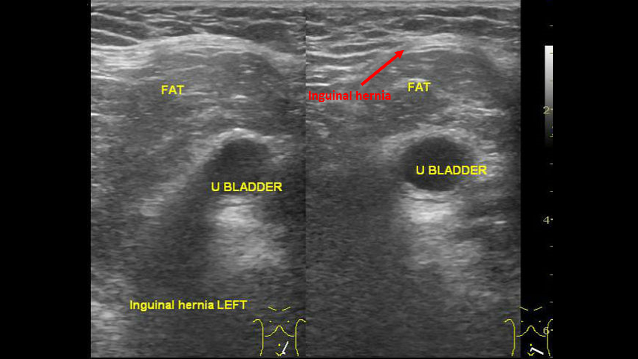
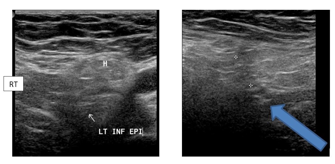
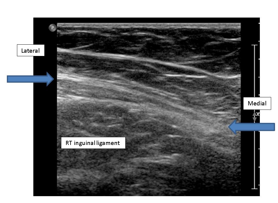
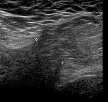
Hernia | UW Ultrasound

BIR Publications
BIR Publications

A) Frontal view of the pelvis passing through the perineal and inguinal...  | Download Scientific Diagram
A) Frontal view of the pelvis passing through the perineal and inguinal... | Download Scientific Diagram

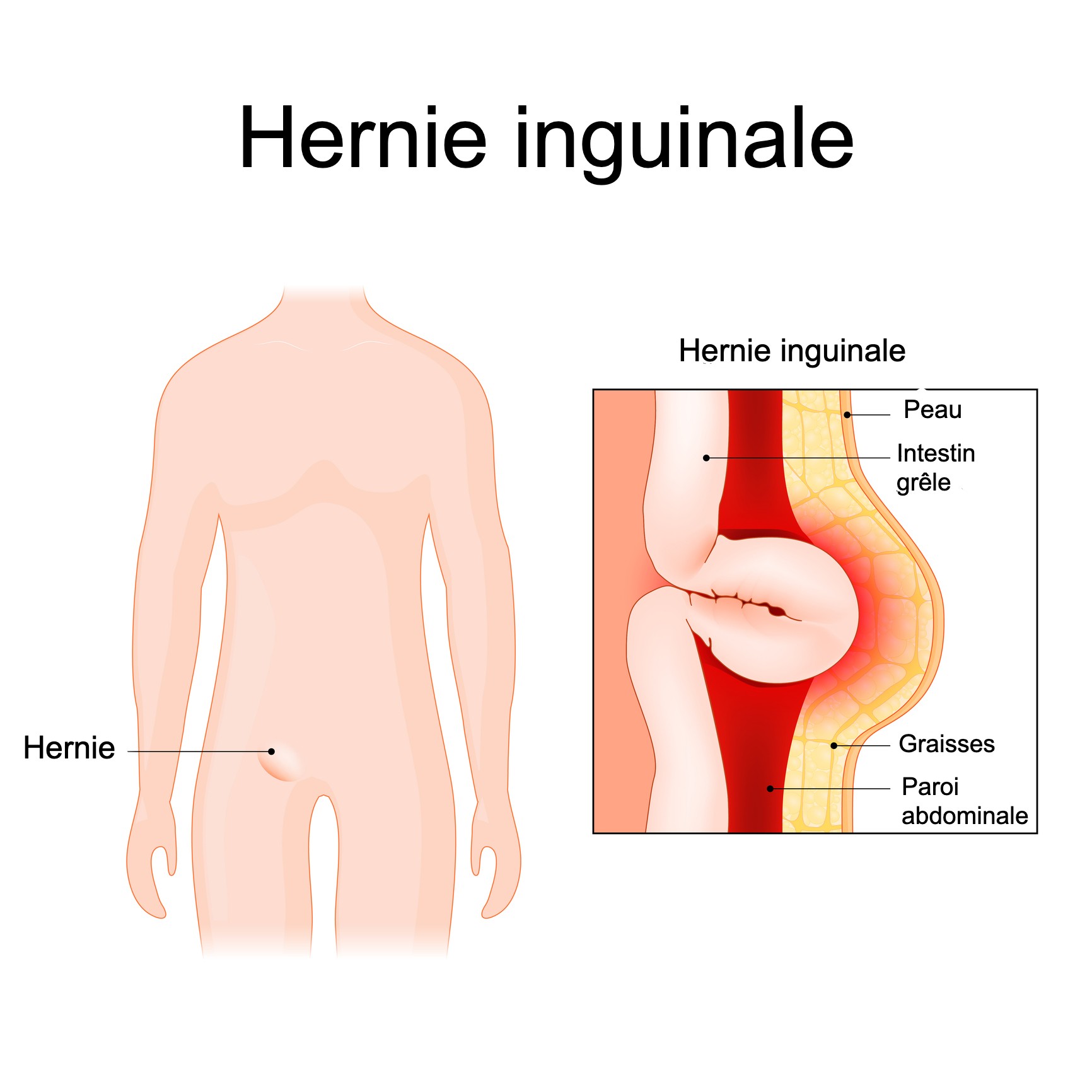
Hernie inguinale - Troubles digestifs - Manuels MSD pour le grand public

View Image
View Image

Undescended testicle cryptorchidism ultrasonography testicule ectopique  échographie ultrasound
Undescended testicle cryptorchidism ultrasonography testicule ectopique échographie ultrasound

Echografie van de inguinale hernia - advanced course MSU - YouTube

Inguinal Hernia in an 8-Year-Old MN Yorkshire Terrier: Our Case Of the  Month: May 2020 | SonoPath
Inguinal Hernia in an 8-Year-Old MN Yorkshire Terrier: Our Case Of the Month: May 2020 | SonoPath

Figure 1 | Ultrasound Prenatal Diagnosis of Inguinal Scrotal Hernia and  Contralateral Hydrocele
Figure 1 | Ultrasound Prenatal Diagnosis of Inguinal Scrotal Hernia and Contralateral Hydrocele

PDF] Hernie inguinale étranglée compliquée d'ischémie testiculaire sur  perméabilité du canal peritonéo-vaginal | Semantic Scholar
PDF] Hernie inguinale étranglée compliquée d'ischémie testiculaire sur perméabilité du canal peritonéo-vaginal | Semantic Scholar

Inguinal hernia ultrasound - wikidoc

Pubalgie: si Hernie inguinale en échographie = Adhésiolyse manuelle  contre-indiquée - YouTube
Pubalgie: si Hernie inguinale en échographie = Adhésiolyse manuelle contre-indiquée - YouTube

Hernia | UW Ultrasound

Figure 3. Ovary-containing hernia of the canal of Nuck in a 1-month-old  girl. A. Longitudinal gray scale ultrasonography shows an ovoid, solid mass  containing cysts in the right inguinal area (arrow), which extends to the  abdominal cavity through the neck of ...
Figure 3. Ovary-containing hernia of the canal of Nuck in a 1-month-old girl. A. Longitudinal gray scale ultrasonography shows an ovoid, solid mass containing cysts in the right inguinal area (arrow), which extends to the abdominal cavity through the neck of ...

BATS - Better Anaesthesia Through Sonography

Hernia | UW Ultrasound

Pathways of Lymphatic Spread in Male Urogenital Pelvic Malignancies |  RadioGraphics
Pathways of Lymphatic Spread in Male Urogenital Pelvic Malignancies | RadioGraphics

Herniated – torsioned ovarian tissue with heterogenous echo structure |  Download Scientific Diagram
Herniated – torsioned ovarian tissue with heterogenous echo structure | Download Scientific Diagram

Hernia | UW Ultrasound

Hernie inguinale : causes, symptômes, opération et traitement - Doctissimo

Inguinal Hernia - Hennepin Healthcare

How to examine the inguinal region | Medmastery

US of the Inguinal Canal: Comprehensive Review of Pathologic Processes with  CT and MR Imaging Correlation
US of the Inguinal Canal: Comprehensive Review of Pathologic Processes with CT and MR Imaging Correlation

Gray-scale ultrasound and power Doppler ultrasound imaging of the right...  | Download Scientific Diagram
Gray-scale ultrasound and power Doppler ultrasound imaging of the right... | Download Scientific Diagram

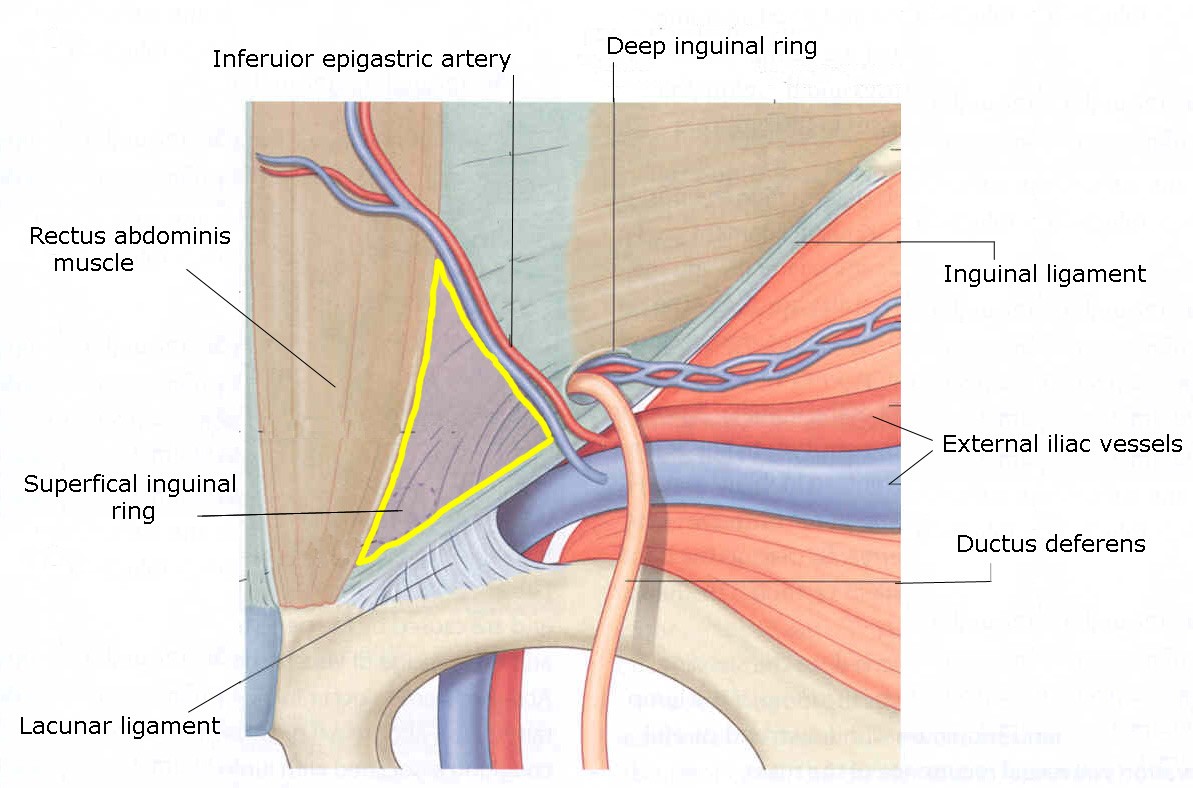
Hernia | UW Ultrasound

A) Inguinal ultrasound depicted a mixed echo mass with intralesional... |  Download Scientific Diagram
A) Inguinal ultrasound depicted a mixed echo mass with intralesional... | Download Scientific Diagram